Torn Retinaculum Knee : Dislocated Kneecap When Is Surgery Recommended - The patella is a sesamoid bone.
byAdmin-
0
Torn Retinaculum Knee : Dislocated Kneecap When Is Surgery Recommended - The patella is a sesamoid bone.. The knee was let to bend down to 90 degrees, and this had excellent repair. The medial collateral ligament (mcl) on the inner side of the knee is most often torn when there is a force that strikes the outside of the knee. When a tear is caused by a medical condition — like tendinitis — the tear usually occurs in the middle of the tendon. Acute tear of medial meniscus of right knee; A new surgical method is introduced offering a less invasive approach to reattach the medial retinaculum following acute patellar dislocation.
The typical injury pattern is a tear of the medial patellofemoral ligament (mpfl) and bone bruises of the patella and the lateral femoral condyle. When a tear is caused by a medical condition — like tendinitis — the tear usually occurs in the middle of the tendon. Upon flexion of the knee, however, a shortened lateral retinaculum will come under excessive stress as the patella is drawn in the trochlea and the iliotibial band pulls posteriorly on the already shortened lateral retinaculum. Current right knee medial meniscus tear; When this happens, you may face a recovery time of weeks to months, depending on the grade of the mcl.
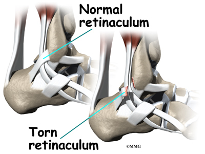
Patellofemoral Pain Syndrome Treatment In Illinois Dr Chams from jkcko2zzf64p2g8m3y9kedlh-wpengine.netdna-ssl.com These may include fractures of the lateral talar process, posterior talar process and anterior process of the calcaneus. Medial means extending toward the middle. Pain on the inside of the knee which may be of sudden onset but can also occur gradually. A complete tear of the patellar tendon indicates a detachment of the kneecap from the shin and is accompanied by an inability to straighten your knee. The lateral patellar retinaculum is a fibrous expansion comprising of superficial and deep layers. The patellar tendon often tears at the place where it attaches to the kneecap, and a piece of bone can break off along with the tendon. Dislocated kneecap, torn medial patellar retinaculum ligament, cartilage, etc. Patellar complications of total knee arthroplasty (tka) are fairly common 1.
The fabellofibular ligament gives minimal supportive strength to the posterolateral corner.
The lateral retinaculum is a ligament that helps hold your patella, or kneecap, in place. The first step in treating a torn meniscus is getting the injury examined by a physician who specializes in orthopedics. The knee was let to bend down to 90 degrees, and this had excellent repair. Despite a decrease in frequency related to improvements in implant design and surgical technique, they still account for about 10% of all tka complications 2. After er doc's saying i must have broken some bone/acl/whatever else, the ortho saying he thought i may have torn my quad tendon, i found out it was none of the above. The surgical technique achieved reinforced reattachment of the torn region of the medial retinaculum for improved patellar support and stabilization. It thickens as it inserts onto the. The ensuing loss of medial restraint favors future patellar dislocations, especially if additional risk factors are present. Pain on the inside of the knee which may be of sudden onset but can also occur gradually. This repaired the quadriceps tendon down anatomically. The typical injury pattern is a tear of the medial patellofemoral ligament (mpfl) and bone bruises of the patella and the lateral femoral condyle. But because of the ligament's location, adequately stretching it can be difficult. The patellar tendon often tears at the place where it attaches to the kneecap, and a piece of bone can break off along with the tendon.
This retrospective analysis comprised 12 cases of medial retinacular repair in 10 patients. It offers an excellent treatment option for people who have experienced more than one dislocation. A complete tear of the patellar tendon indicates a detachment of the kneecap from the shin and is accompanied by an inability to straighten your knee. The medial patellar retinaculum is a tendon of the knee that crosses the knee joint on the medial side of the patella. The fabellofibular ligament gives minimal supportive strength to the posterolateral corner.
Peroneal Tendon Subluxation Eorthopod Com from eorthopod.com Additionally, complex injuries to bone, cartilage, and ligaments may occur. The superficial layer originates from the lowest fibers of the iliotibial band and from an extension of vastus lateralis fascia. The patellar tendon often tears at the place where it attaches to the kneecap, and a piece of bone can break off along with the tendon. A complete tear of the patellar tendon indicates a detachment of the kneecap from the shin and is accompanied by an inability to straighten your knee. A complete tear of the patellar tendon. After er doc's saying i must have broken some bone/acl/whatever else, the ortho saying he thought i may have torn my quad tendon, i found out it was none of the above. It inserts onto the medial aspect of the patellar. The medial patellar retinaculum is a tendon of the knee that crosses the knee joint on the medial side of the patella.
The typical injury pattern is a tear of the medial patellofemoral ligament (mpfl) and bone bruises of the patella and the lateral femoral condyle.
Stephen pribut's running injuries website. Partial patellectomy 3 moths ago.pain now above the knee.mri results show hoff fluid spoiled,torn lat.retinaculum,muscle fluid. It inserts onto the medial aspect of the patellar. Stretching this ligament keeps the patella in place and the ligament healthy. But because of the ligament's location, adequately stretching it can be difficult. The lateral knee retinaculum is oriented longitudinally with the knee extended. The superficial layer originates from the lowest fibers of the iliotibial band and from an extension of vastus lateralis fascia. We put the knee in extension and tied down the sutures. The medial collateral ligament (mcl) on the inner side of the knee is most often torn when there is a force that strikes the outside of the knee. The lateral retinaculum is a ligament that helps hold your patella, or kneecap, in place. The fabellofibular ligament gives minimal supportive strength to the posterolateral corner. A complete tear of the patellar tendon. These may include fractures of the lateral talar process, posterior talar process and anterior process of the calcaneus.
They are minor patellar stabilizers and, if intact, can provide knee extension and straight leg raising despite a patellar or quadriceps tendon rupture. The medial and lateral patellar retinaculum are on their respective sides of the patella and are continuous with the vastus fascia to the tibia and the patella. The medial retinaculum plays a minor role — along with the vastus medialis oblique and the medial patellofemoral ligament — in providing medial stability in the knee, according to dr. Peter ihle answered 54 years experience orthopedic surgery It inserts onto the medial aspect of the patellar.
Kjr Korean Journal Of Radiology from www.kjronline.org the mcl attempts to resist the knee bending sideways and tears if the force is too great. Stephen pribut's running injuries website. Pain on the inside of the knee which may be of sudden onset but can also occur gradually. The medial and lateral patellar retinaculum are on their respective sides of the patella and are continuous with the vastus fascia to the tibia and the patella. Patellar contusions and avulsion fractures arealsoidentified inupto41% ofpatients i121.thepatellar injuries areadjacent tothe attachment ofthemedial retinaculum and tend toinvolve themedial andinferior aspects ofthebone. The typical injury pattern is a tear of the medial patellofemoral ligament (mpfl) and bone bruises of the patella and the lateral femoral condyle. A complete tear of the patellar tendon indicates a detachment of the kneecap from the shin and is accompanied by an inability to straighten your knee. The procedure is relatively new.
These may include fractures of the lateral talar process, posterior talar process and anterior process of the calcaneus.
1 doctor answer • 1 doctor weighed in. It inserts onto the medial aspect of the patellar. In view of this, there is little to support the lateral tibial plateau posteriorly once the anterior cruciate ligament has been torn. Patellar contusions and avulsion fractures arealsoidentified inupto41% ofpatients i121.thepatellar injuries areadjacent tothe attachment ofthemedial retinaculum and tend toinvolve themedial andinferior aspects ofthebone. The sutures were then cut. This medical condition requires immediate intervention from an orthopedic surgeon, as sometimes a piece of the patella bone may break off along with the tendon and cannot reattach on its own. Additionally, complex injuries to bone, cartilage, and ligaments may occur. Mri show obliq tear body and posterior horn lateral meniscus, extending infr artic surface and ulceration articular cartilage patella. This occurs because of the chronic pull of the knee cap to the outside by the thigh muscles, creating a strain on the medical or inside tissues (the retinaculum). The medial patellar retinaculum is a tendon of the knee that crosses the knee joint on the medial side of the patella. These may include fractures of the lateral talar process, posterior talar process and anterior process of the calcaneus. The superficial layer originates from the lowest fibers of the vastus medialis muscle, sartorius and the medial collateral ligament.the deep layer has contributions from the medial patellofemoral ligament and fascial thickenings. We put the knee in extension and tied down the sutures.